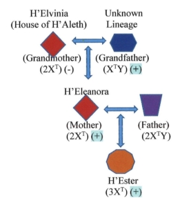
H’Ester agreed to have her DNA analyzed at the natural history and research center for the number and shape of chromosomes in her cells and the location of genes along individual chromosomes. Master Mikalov advised Matron Aquina that the karyotype could be done by the next day. The remainder of the requested analyses would have to wait. They would be put in the queue along with all the other analyses the research center had put on hold. Their top priority was conducting an analysis of the genomic structure of the lyvulfon from the tissue sample Master Xavier had brought back. By the next day, the center’s staff had two top priorities. H’Ester did not have two XT chromosomes. She had three. (Excerpt from Homo transformans: The Origin and Nature of the Species.)
Introduction
Humans typically have 23 pairs of chromosomes in every cell for a total count of 46 in each cell. The reproductive chromosomes, X and Y, are one of these pairs. All nonreproductive cells should have a full complement of 46 chromosomes. Reproductive cells ‒ an ovum (egg) or a sperm ‒ should have only one copy of each chromosome, including either an X or a Y chromosome, for a total of 23 single chromosomes. Once an ovum and a sperm merge, then the resulting fertilized ovum will have a full complement of 46 chromosomes, including either a pair of XX chromosomes for a female or a pair of XY chromosomes for a male. Yet, this is not always the case.
Aneuploidy
Aneuploidy is an abnormal number of chromosomes (Jorde, 2014). In humans, this is reflected in cells that have either fewer than or more than 46 chromosomes. If someone has an extra chromosome for a total of 47 chromosomes, then the individual has a genetic condition known as trisomy.
Trisomy
March is Trisomy Awareness month. Trisomy occurs when a chromosome pair fails to separate properly during development of an ovum or a sperm (MacLennon, et al., 2015). When that happens, one ovum or sperm will receive both copies of that chromosome while its twin ovum or sperm receives no copy. The presence of an additional chromosome over the normal complement of 46 is not uncommon. Depending upon which chromosome has an extra third member, the fetus may continue to develop and result in the birth of a baby. The redundant chromosome, however, could lead to developmental disabilities that may or may not become apparent.
One of the most common examples of trisomy in a nonreproductive chromosome is Downs Syndrome (trisomy 21). In this disorder, there are three copies of chromosome 21 instead of the customary two copies. This is usually a congenital abnormality associated with increased maternal age, in which case it is not an inherited disorder.
The most commonly encountered examples of an extra reproductive chromosome involves duplication of the X chromosome. These include Trisomy X (47, XXX) in female offspring, (Otter, et al., 2010; Tartaglia, et al., 2010) and Kleinfelter’s syndrome (47, XXY) in male offspring (Skuse, et al., 2018; Visootsak, et al., 2006). Both males and females may have physical and mental developmental disabilities; however, their disabilities may be quite subtle. Hence, many people with Trisomy X and Kleinfelter’s syndrome may go unrecognized.
Additional information about trisomy and other chromosome disorders can be found at https://ghr.nlm.nih.gov/search?query=trisomy. Information about genes and chromosomes in general can be found at https://ghr.nlm.nih.gov/gene and https://ghr.nlm.nih.gov/chromosome, respectively.
References
Jorde, L. B. (2014.) Genes and genetic diseases. In: K. L. McCance, S. E. Huether, V. L. Brashers, V.L., and N. S. Rote, Pathophysiology: The Biologic Basis for Disease in Adults and Children (7th ed.), Chapter 4. St. Louis: Mosby/Elsevier.
MacLennan, M., Crichton, J. H., Playfoot, C. J., & Adams, I. R. (2015.) Oocyte development, meiosis and aneuploidy. Seminars in cell & developmental biology, 45, 68–76. https://doi.org/10.1016/j.semcdb.2015.10.005.
Otter, M., Schrander-Stumpel, C. T., Curfs, L. M. (2010.) Triple X syndrome: a review of the literature. Eur J Hum Genet, 18(3), 265-71. doi: 10.1038/ejhg.2009.109.
Skuse, D., Printzlau, F., Wolstencroft, J. (2018.) Sex chromosome aneuploidies. Handb Clin Neurol, 147, 355-376. doi: 10.1016/B978-0-444-63233-3.00024-5.
Tartaglia, N. R., Howell, S., Sutherland, A., et al. (2010.) A review of trisomy X (47,XXX). Orphanet J Rare Dis, 11, 5, 8. doi: 10.1186/1750-1172-5-8.
Visootsak, J., and Graham, J. M. Jr. (2006.) Klinefelter syndrome and other sex chromosomal aneuploidies. Orphanet J Rare Dis, 24 (1), 42. doi: 10.1186/1750-1172-1-42.